Home
/ Anatomy Of Musckes Sndctendons / Ankle Anatomy Be In Motion Physiotherapy : There's no strict demarcation or dividing line between the tendon and the covering around this muscle but that covering is called is called the epimysium fp my cm and it's really just connective tissue that covers the muscle kind of protects it reduces friction.
Anatomy Of Musckes Sndctendons / Ankle Anatomy Be In Motion Physiotherapy : There's no strict demarcation or dividing line between the tendon and the covering around this muscle but that covering is called is called the epimysium fp my cm and it's really just connective tissue that covers the muscle kind of protects it reduces friction.
Anatomy Of Musckes Sndctendons / Ankle Anatomy Be In Motion Physiotherapy : There's no strict demarcation or dividing line between the tendon and the covering around this muscle but that covering is called is called the epimysium fp my cm and it's really just connective tissue that covers the muscle kind of protects it reduces friction.. The anatomy of muscle cells differs from that of other body cells and biologists have applied specific terminology to different parts of these cells. The muscular system consists of the skeletal muscles and their associated structures. Circular skeletal muscles are made up of fibers that are arranged in a circular manner. Muscular contraction is necessary for voluntary and involuntary movement of limbs, stabilization of joints, maintaining luminal diameter (in the case of arteries, bowel, etc), and to produce heat. The muscles of the torso, examined in the previous chapter, include a few that attach directly into the upper arm and help move the humerus at the shoulder joint.
How to study muscle anatomy. An interactive tutorial teaching the position, actions, innervation and attachments of the rectus femoris muscle with the aid of anatomical illustrations. Discover the muscle anatomy of every muscle group in the human body. Learn about human anatomy muscles with free interactive flashcards. Anatomy of the muscular system.
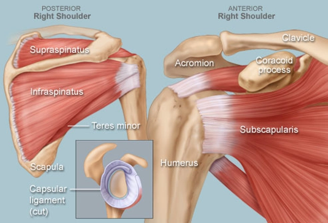
Shoulder Human Anatomy Image Function Parts And More from img.webmd.com The smooth muscle tissue that forms organs like the stomach and bladder changes. Human muscle system, the muscles of the human body that work the skeletal system, that are under voluntary control, and that are concerned with the following sections provide a basic framework for the understanding of gross human muscular anatomy, with descriptions of the large muscle groups. In the diagrams below, i'll be showing muscle groups in color, with a black line to show the forms that would show through the skin (i also show protruding bones that would do the same). Muscular contraction is necessary for voluntary and involuntary movement of limbs, stabilization of joints, maintaining luminal diameter (in the case of arteries, bowel, etc), and to produce heat. Along with lateral pterygoid muscle it produces side to side movement of mandible. Skeletal muscles are attached to bones by tendons and can be as long as 30 cm, although they are usually 2 to 3 cm in length. Smooth muscles are found in the walls of many organs, such as the stomach and in blood vessels. Discover the muscle anatomy of every muscle group in the human body.
Anatomy of the short head of the biceps brachii muscle.
As the skeletal muscles pull on bones to cause movements, they also stabilize the joints of the skeleton; Cardiac muscle contracts the heart to pump blood. The anterior and middle scalenes originate from the transverse processes of certain cervical vertebrae and attach to the first rib. The muscles around the knee help to keep the knee stable, well aligned, and moving. Related online courses on physioplus. From anterior to posterior, the tongue has 3 surfaces: The tip is the highly mobile, pointed anterior portion of the tongue. Adducts & flexes the arm (humerus). There's no strict demarcation or dividing line between the tendon and the covering around this muscle but that covering is called is called the epimysium fp my cm and it's really just connective tissue that covers the muscle kind of protects it reduces friction. Anatomy of the short head of the biceps brachii muscle. See the pictures and anatomy description of knee joint bones, cartilage, ligaments, muscle and tendons with resources for knee problems & injuries. There are two main muscle groups around the knee: In the diagrams below, i'll be showing muscle groups in color, with a black line to show the forms that would show through the skin (i also show protruding bones that would do the same).
However, if you take a little time to learn a few root words, those latin. This is a table of skeletal muscles of the human anatomy. Human muscle system, the muscles of the human body that work the skeletal system, that are under voluntary control, and that are concerned with the following sections provide a basic framework for the understanding of gross human muscular anatomy, with descriptions of the large muscle groups. General functions of muscular system: Muscular contraction is necessary for voluntary and involuntary movement of limbs, stabilization of joints, maintaining luminal diameter (in the case of arteries, bowel, etc), and to produce heat.
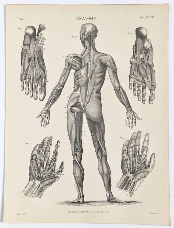
1890s Antique Anatomy Print Muscles And Tendons Human Body Etsy from i.etsystatic.com Microscopic anatomy of skeletal muscle. Related online courses on physioplus. The muscular system consists of the skeletal muscles and their associated structures. In the diagrams below, i'll be showing muscle groups in color, with a black line to show the forms that would show through the skin (i also show protruding bones that would do the same). Anatomy of the short head of the biceps brachii muscle. Attached to the bones of the skeletal system are about 700 named muscles that make up roughly half of a person's body weight. Along with lateral pterygoid muscle it produces side to side movement of mandible. Skeletal muscle moves bones and other structures.
Each of these muscles is a discrete organ constructed of skeletal muscle tissue, blood vessels, tendons, and nerves.
Skeletal muscles are attached to bones by tendons and can be as long as 30 cm, although they are usually 2 to 3 cm in length. This is a table of skeletal muscles of the human anatomy. Learn about human anatomy muscles with free interactive flashcards. Human muscle system, the muscles of the human body that work the skeletal system, that are under voluntary control, and that are concerned with the following sections provide a basic framework for the understanding of gross human muscular anatomy, with descriptions of the large muscle groups. Anatomy, function, and rehab considerations. There are around 650 skeletal muscles within the typical human body. Skeletal muscles allow the body to move and maintain posture; The three scalene muscles are found forming the floor of the posterior triangle. The anatomy of muscle cells differs from that of other body cells and biologists have applied specific terminology to different parts of these cells. Attached to the bones of the skeletal system are about 700 named muscles that make up roughly half of a person's body weight. Learn more about how muscles work, what they look like, and how they're treated. The tendons of these muscles pass through a small corridor in the wrist known as the carpal tunnel. As the skeletal muscles pull on bones to cause movements, they also stabilize the joints of the skeleton;
As the skeletal muscles pull on bones to cause movements, they also stabilize the joints of the skeleton; Muscular contraction is necessary for voluntary and involuntary movement of limbs, stabilization of joints, maintaining luminal diameter (in the case of arteries, bowel, etc), and to produce heat. Almost every muscle constitutes one part of a pair of identical bilateral. Learning to draw muscles may conjure medical charts in daunting details, but such complexity is unnecessary. The muscular system is responsible for the movement of the human body.
Quadriceps Tendonitis Richmond Va Knee Surgery Richmond from www.kneeandshouldersurgery.com See the pictures and anatomy description of knee joint bones, cartilage, ligaments, muscle and tendons with resources for knee problems & injuries. From anterior to posterior, the tongue has 3 surfaces: The muscles around the knee help to keep the knee stable, well aligned, and moving. Related online courses on physioplus. The muscular system consists of about 700 muscle organs that are typically attached to bones across a joint to produce all voluntary movements. Learn more about how muscles work, what they look like, and how they're treated. Learn about human anatomy muscles with free interactive flashcards. The three scalene muscles are found forming the floor of the posterior triangle.
A collection of anatomy notes covering the key anatomy concepts that medical students need to learn.
Adducts & flexes the arm (humerus). The tendons of these muscles pass through a small corridor in the wrist known as the carpal tunnel. Learn about human anatomy muscles with free interactive flashcards. This article will focus on tongue embryology, origin, insertion, and action of the extrinsic muscles, followed by innervation, blood supply and lymphatic drainage of the tongue. Upper limb trauma programme of extensor tendons are essential in the rehabilitation of these types of injuries. Muscular contraction is necessary for voluntary and involuntary movement of limbs, stabilization of joints, maintaining luminal diameter (in the case of arteries, bowel, etc), and to produce heat. The three scalene muscles are found forming the floor of the posterior triangle. In the muscular system, muscle tissue is categorized into three distinct types: Muscles of mastication are classified as main and accessory muscles. However, if you take a little time to learn a few root words, those latin. It occupies most of the oral cavity and oropharynx. Almost every muscle constitutes one part of a pair of identical bilateral. There are around 650 skeletal muscles within the typical human body.